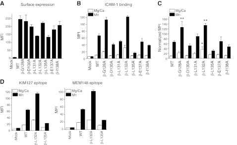
Figure 5.
Functional role of the α′1 helix of the βI domain. A) Surface expression of alanine-scanning mutant in the α′1 helix probed by TS2/4 antibody. MFI; mean fluorescence intensity. B) ICAM-1 binding of mutants in 1 mM Mg2+/Ca2+ or 1 mM Mn2+. C) ICAM-1 binding of mutants normalized with surface expression. Normalized MFI of ICAM-1 binding was calculated as [MFI of ICAM-1 binding of mutant] × {[(MFI of surface expression of WT) − (MFI of surface expression of mock transfectant)]/[(MFI of surface expression of mutant) − (MFI of surface expression of mock transfectant)]}. One-way ANOVA with Tukey post hoc was performed to compare data within Mg2+/Ca2+ or Mn2+ group. *P < 0.05 vs. WT Mg2+/Ca2+, **P < 0.05 vs. WT Mn2+. D) Leg extension and headpiece opening in β-L132A and β-L135A mutants probed by KIM127 and MEM148 antibodies, respectively. Data are shown as means ± se of 3 independent experiments of triplicates.

