Figure 4.

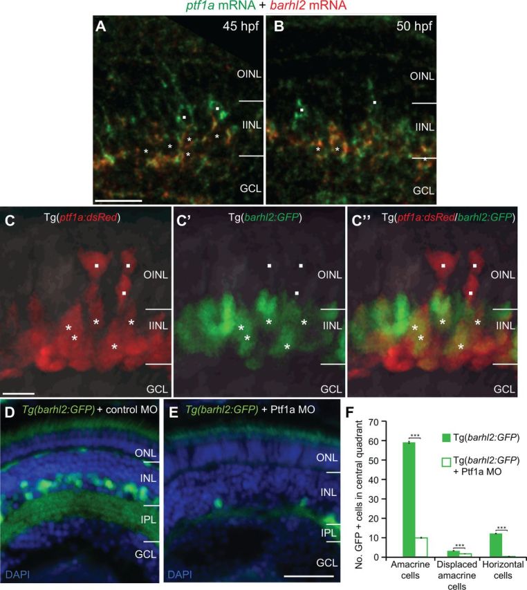
Ptf1a/barhl2 are sequentially expressed within individual cells, with Ptf1a being necessary for Barhl2:GFP expression. A, B, Double fluorescent in situ hybridization of barhl2 and ptf1a mRNAs. Ptf1a (green) is expressed in cells apically (squares) and in cells that have migrated basally where they also express barhl2 (red, asterisks) at 45 hpf (A) and 50 hpf (B). C–C″, Micrographs from in vivo time-lapse of double transgenic Tg(ptf1a:dsRed/barhl2:GFP) embryos at 35 hpf show a similar pattern with apical cells expressing Ptf1a:DsRed alone (squares) and more basal cells coexpressing Barhl2:GFP (asterisks). D, E, Micrographs of 120 hpf Tg(barhl2:GFP) injected with standard morpholino (D) or Ptf1a morpholino (E). Barhl2:GFP expression is drastically reduced in the Ptf1a morphants. F, Quantification of GFP-labeled cells shows a significant loss of cells in Ptf1a MO in all cell types that usually express Barhl2:GFP, i.e., ACs (inner half of INL), displaced ACs (outermost layer of GCL) and horizontal cells (outermost layer of INL). WT n = 51 eyes, Ptf1a morphant n = 41 eyes. ONL, Outer nuclear layer; IPL, inner plexiform layer; GCL, ganglion cell layer. ***p < 0.001; error bars indicate SEM. Scale bars: (in A) A, B, 20 μm; (in C) C–C″, 10 μm; (in E) D, E, 50 μm.
