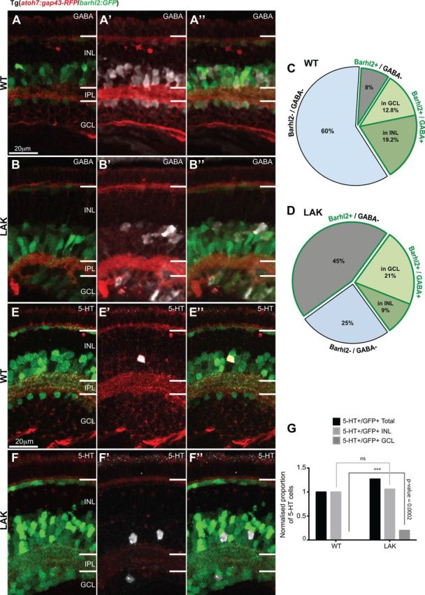
Figure 9.

Absence of Atoh7 leads to an increase of barhl2-expressing ACs in the ganglion cell layer. A, B, Micrographs of 120 hpf Tg(atoh7:gap43-RFP/barhl2:GFP) retinas labeled for GABA in WT (A) and lakritz (LAK) (B). C, D, Quantification of amacrine subpopulations expressed as proportion of total INL and GCL cells. The data indicate that the increase in the number of GFP+ cells in the LAK retina is primarily due to non-GABAergic ACs, as the proportion of GABAergic ACs remains unchanged in total in both GCL and INL in LAK versus WT (32% WT to 30% lakritz). A redistribution of GABAergic ACs is, however, observed with an increase of Barhl2+/GABA+ cells in the GCL and a relative decrease of those cells in the INL of the LAK retina compared with WT (12.8% WT to 21% LAK, p = 0.0008 in GCL and 19.2% WT to 9% LAK, p = 0.0014 in INL). Total number of cells is the number of nuclei in the INL and GCL. E, F, Micrographs of 120 hpf Tg(atoh7:gap43-RFP/barhl2:GFP) retinas labeled for serotonin (5-HT). G, Quantification of the total proportion of serotonin-positive/Barhl2:GFP-positive (5-HT+/GFP+) neurons shows an increase in 5-HT cells in lakritz specifically in the GCL where they are normally absent (n = 15–19 eyes). IPL, Inner plexiform layer; GCL, ganglion cell layer. ***p < 0.0002. Error bars indicate SEM. Scale bars, 20 μm.
