Figure 5.
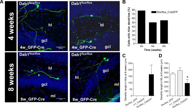
Dab1 knock-out adult-generated neurons exhibit ectopic basal dendrites in the hilus. A, Representative images of Dab1flox/flox neurons infected with GFP and Cre, with hilar dendrites at 4 and 8 weeks after surgery. hl, Hilus; gcl, granule cell layer; ml, molecular layer. B, Between 50% and 78% of adult-generated cells in Dab1flox/flox neurons infected with Cre exhibited hilar dendrites. This observation contrasts with the two control groups, in which no hilar dendrites were detected. C, Presence of dendritic arborization in the hilus of Cre-infected Dab1flox/flox neurons while this was absent in the other groups. D, The dendritic arborization in the ML of Cre-infected Dab1flox/flox neurons was lower than in control groups (ANOVA: p < 0.05; post hoc: p < 0.05).

