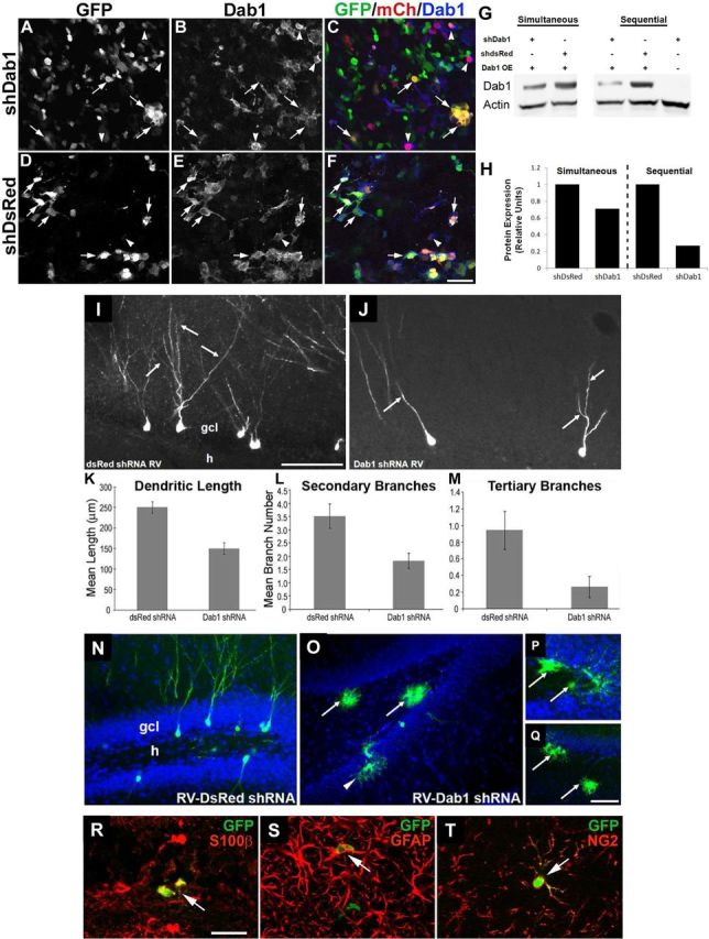
Figure 6.

Less complex dendritic arbors form on adult-generated DGCs in rat after shRNA-induced Dab1 suppression. A–F, Cultured 293T cells were cotransfected with Dab1 overexpression vector (pDab1-IRES-mCh-WPRE) and either pSiE-shDab1-EF1α-GFP (A–C) or pSiE-shDsRed-EF1α-GFP (D–F), and 48 h later were fixed and triple labeled for GFP, mCh, and Dab1. Yellow cells coexpressing mCh and GFP have lower Dab1 expression after knockdown with shDab1 (A–C, arrows) compared with similar cells in control cultures (D–F, arrows). Arrowheads in A–F denote cells with strong Dab1 expression in the setting of low or absent GFP. Note that mCh expression was lower in the shDsRed-expressing cultures (compare F with C). Scale bar: (in F) A–F, 25 μm. G, H, Immunoblot (G) and densitometric quantification (H) of Dab1 protein expression in cultured 293T cells 48 h after transfection with varying combinations of Dab1 overexpression (OE) vector and shDab1 or shDsRed. Transfections were either performed simultaneously or with RNAi vector followed 6 h later by Dab1 OE vector (sequential). Sequential transfection of Dab1shRNA and then Dab1OE vector resulted in a >70% decrease in Dab1 protein levels compared with the control condition (G, lanes 4 and 5; H, right-sided bars). I, J, RV-mediated expression of Dab1 shRNA in DGC progenitors markedly decreased dendritic arborization of GFP-labeled cells in the granule cell layer (gcl) of an adult rat (J, arrows) compared with control DsRed shRNA-infected DGCs (I, arrows). RV shRNA vectors were injected 4 weeks earlier. Scale bar: (in I) I, J, 50 μm. K–M, Quantification of dendritic complexity showed that Dab1 shRNA-expressing DGCs had decreased mean dendritic length (K, p < 0.001), and fewer secondary (L, p < 0.005) and tertiary branches (M, p < 0.05). N–Q, Representative confocal images of adult rat DG 4 weeks after control RV-U6-shDsRed-EF1α-GFP injection (N) shows GFP-labeled DGCs with a normal appearance and no labeled glia. In contrast, after RV-U6-shDab1-EF1α-GFP injection, many labeled cells with glial morphology were evident in the DG (O–Q). Arrows indicate putative glial cells in the hilus (h), and the arrowhead in O denotes another in the gcl. Scale bar: (in Q) O, Q, 50 μm; (in Q) N, P, 25 μm. R–T, Confocal images of sections through the DG of adult rats 4 weeks after injection of RV-U6-shDab1-EF1α-GFP double-immunolabeled for GFP (green) and S100β (R, red), GFAP (S, red), or NG2 (T, red). Arrows denote double-labeled cells in the dentate hilus. Scale bar: R, 25 μm.
