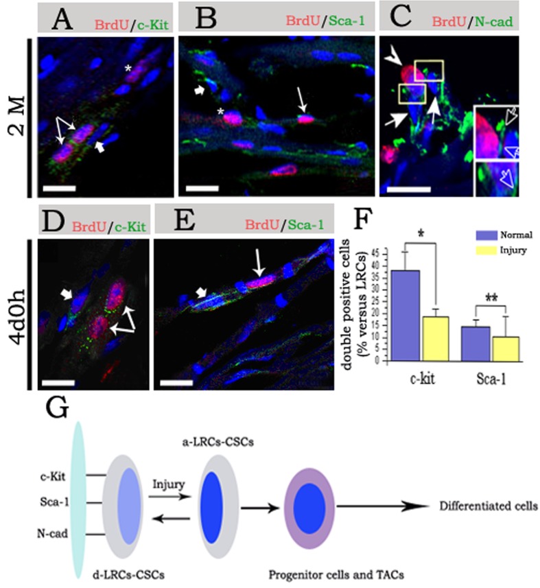
Figure 4. Molecular characteristics of CSCs/CPCs during chasing and post-injury.
(A)–(C) Show staining of cardiac tissues in 2 months chasing. (A) indicates staining of BrdU and c-Kit, the 2 fine arrows point to double positive cells, and the asterisk indicates BrdU+/c-Kit− cells (asterisk); however, the thick arrow points to BrdU−/c-Kit+ cells (arrow). (B) Indicates staining of BrdU and Sca-1, a fine arrow points to double positive cells, and the asterisk indicates BrdU+ and Sca-1− cells; however, the thick arrow points to BrdU−/Sca-1+ cells (arrow). (C) Indicates staining of BrdU and N-cad, the arrowhead indicates BrdU+/N-cad+ cells adhering to BrdU−/N-cad+ spindle cells (arrow), the low right corner in (C) are magnification of rectangular region, and the hollow arrow indicate N-cad expression between the BrdU+ cells and BrdU− cells. (D and E) Show staining of cardiac tissues at day 4 post-injury; (D) indicates staining of BrdU and c-Kit. The 2 fine arrow point to double positive cells; however, the thick arrow points to BrdU−/c-Kit+ cells (arrow). (E) Indicates staining of BrdU and Sca-1, fine arrow point to double positive cells, however, the thick arrow points to BrdU−/Sca-1+ cells (arrow). Nuclei were staining with DAPI. (F) Shows statistical of double positive cells. *P<0.01 show most significance and **P>0.05 shows no significance. (G) Shows a dynamic model of CSCs/CPCs regeneration post-injury. The d-CSCs/CPCs pool (d-LRC-CSCs/CPCs), circulate only a few times over the life span of the mouse in a homoeostatic situation, but is activated (a-LRC-CSCs/CPCs) and participated in replenishment of the cardiac tissues post-injury (scale bars: A, B, D and E = 20 μm; C = 10 μm).

