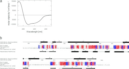
Figure 3. Structural analyses of sAC-HD by UV–CD spectroscopy and structural comparison to haem-containing PAS domains.
(A) UV–CD spectrum indicating a secondary structure content of 34% α-helix and 30% β-sheet. The molar elipticities given are relative to the mean amino acid weight. (B) Alignment of sAC-HD and the haem-containing PAS domains and PAC domains of FixL-RHIME (Rhizobium meliloti) and FixL-BRAJA (Bradyrhizobium japonicum). Blue coloured residues indicate high conservation of physicochemical properties and red colour reflects medium conservation. The secondary structure prediction for sAC-HD on top of the alignment was generated with Jpred [48] and the representative PAS-fold shown below the alignment was deduced from a structural alignment of six PAS structures [33].

