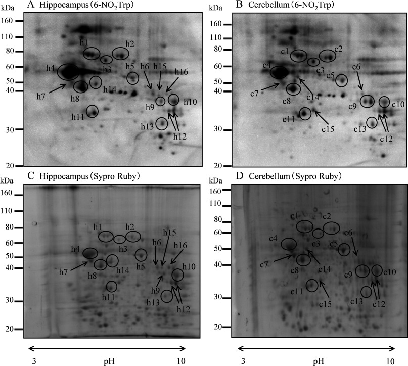
Figure 1. Representative results of Western blotting with an anti-6-NO2Trp antibody and Sypro Ruby-stained gel after two-dimensional electrophoresis.
(A) and (C) are Western blotting and Sypro Ruby gel staining of hippocampus respectively. (B) and (D) are those of cerebellum. Open circled spots and spots indicated by arrows in hippocampus (h1–h16) and cerebellum (c1–c15) were subjected to LC-ESI-MS/MS analysis.

