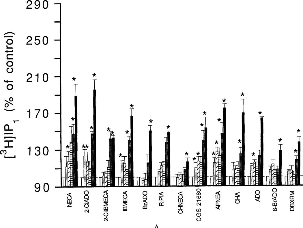
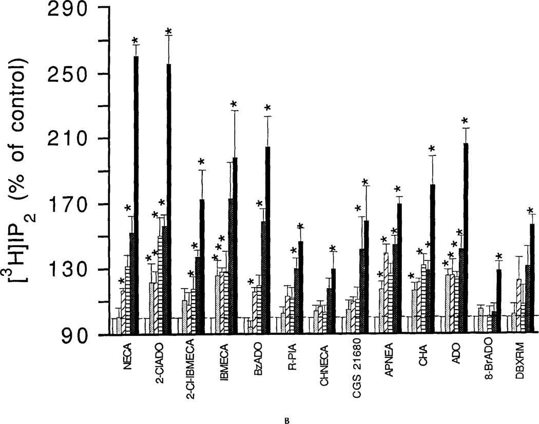
Figure 1.
Concentration-dependent stimulation of phosphoinositide breakdown by adenosine analogs in RBL-2H3 mast cells. A: [3H]IP1. B: [3H]IP2. Accumulation of [3H]inositol phosphates was measured as described in Methods in the absence or presence of adenosine analogs. Analogs were as follows: NECA, 2-chloroadenosine (2CIADO); 2Cl-IBMECA; N6-3-iodobenzyl-MECA (IBMECA); N6-benzyladenosine (BzADO); N6-R-phenylisopropyladenosine (R-PIA); N6-cyclohexylNECA (CHNECA); CGS 21680; APNEA; N6-cyclohexyladenosine (CHA); adenosine (ADO); 8-bromoadenosine (8-BrADO); 1,3-dibutylxanthine 7-riboside-5′-N-methylcarboxamide (DBXRM). The analogs were tested in the concentration range of 0.3 to 30 µM (open bars control; dotted bars 0.3 µM; hatched bar 1 µM; striped bar 3 µM; heavy dotted bar 10 µM; solid bar 30 µM). Incubations were for 1 min with cells pretreated with the DNP-specific IGE and then challenged with the antigen DNP-HSA. Basal accumulations of [3H]IP1 and [3H]IP2 for the 1-min incubations were 258 ± 21 and 143 ± 10 cpm/10,000 cpm membrane lipids, respectively. Values are means ± s.e.m. (n = 3). *p <0.05.


