
Figure 3.
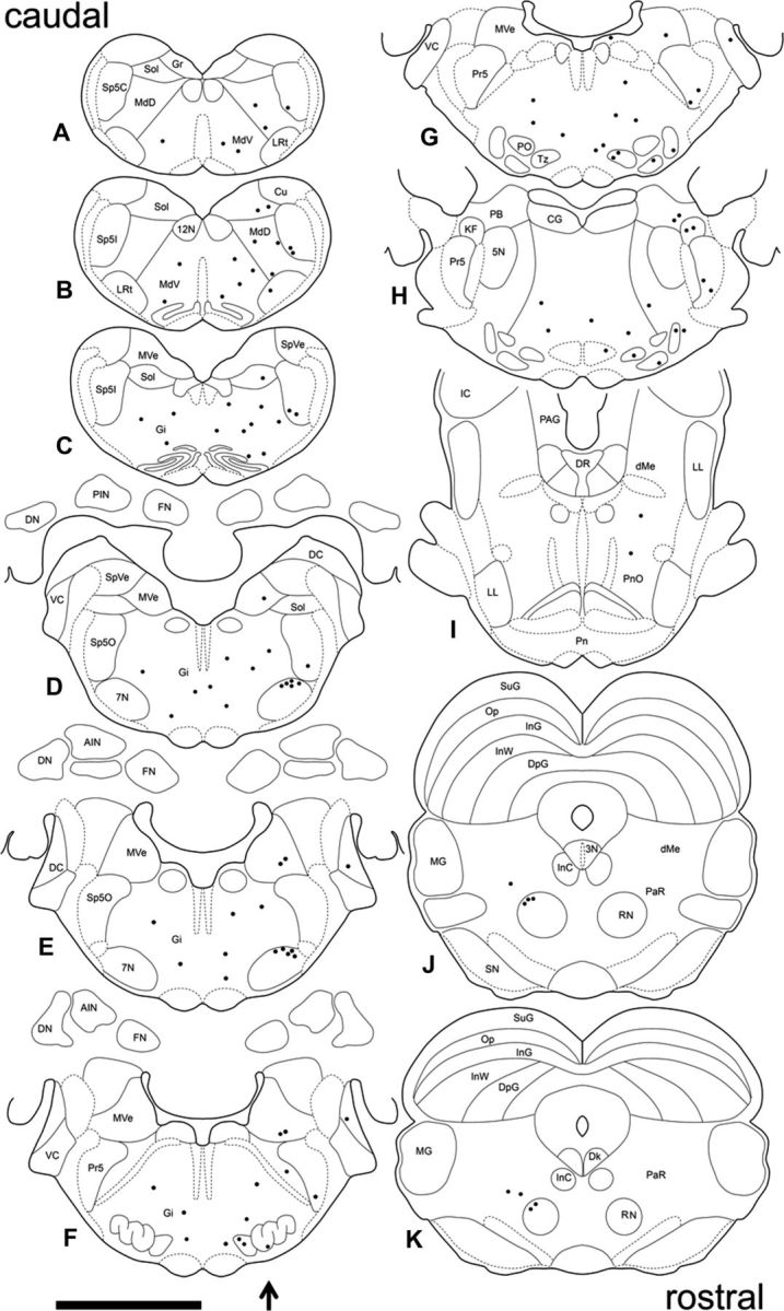
Distribution of PRV-labeled neurons in the brain 4 d after PRV injection in the right OOM. A composite of the location of PRV-labeled neurons found across four animals plotted in sequential sections to best represent the pattern of PRV labeling. Sections are arranged caudorostrally (A–K). The arrow indicates the side of PRV injection. Each dot represents one PRV-labeled neuron. 3N, Oculomotor nucleus; 4N, trochlear nucleus; 5N, motor trigeminal nucleus; 7N, motor facial nucleus; 12N, hypoglossal nucleus; AIN, anterior interpositus; CG, central gray; Cn, cuneiform nucleus; DC, dorsal cochlear; Dk, nucleus of Darkschewitsch; LL, lateral lemniscus; dMe, deep mesencephalic nucleus; DN, dentate nucleus; PAG, periaqueductal gray; DpG, deep layer of the superior colliculus; FN, fastigial nucleus; Gi, gigantocellular reticular nucleus; Gr, gracile nucleus; IC, inferior colliculus; InC, interstitial nucleus of Cajal; InG, intermediate gray layer of the superior colliculus; InW, intermediate white layer of the superior colliculus; KF, Kölliker-fuse nucleus; LRt, lateral reticular nucleus; MdD, dorsal medullary reticular nucleus; MdV, ventral medullary reticular nucleus; MG, medial geniculate nucleus; MVe, medial vestibular nucleus; Op, optic nerve layer of the superior colliculus; PaR, pararubral nucleus; PB, parabrachial nucleus; Pn, pontine nucleus; PnO, pontine reticular nucleus, oral; PO, periolivary nucleus; Pr5, principal sensory trigeminal nucleus; RN, red nucleus; RRF, retrorubral field; SN, substantial nigra; Sol, solitary nucleus; Sp5C, spinal trigeminal nucleus, caudal; Sp5I, spinal trigeminal nucleus, interpolar; Sp5O, spinal trigeminal nucleus, oral; SpVe, spinal vestibular nucleus; SuG, superficial gray layer of the superior colliculus; tth, trigeminothalamic tract; Tz, nucleus of the trapezoid body; VC, ventral cochlear nucleus. Scale bar, 5 mm.