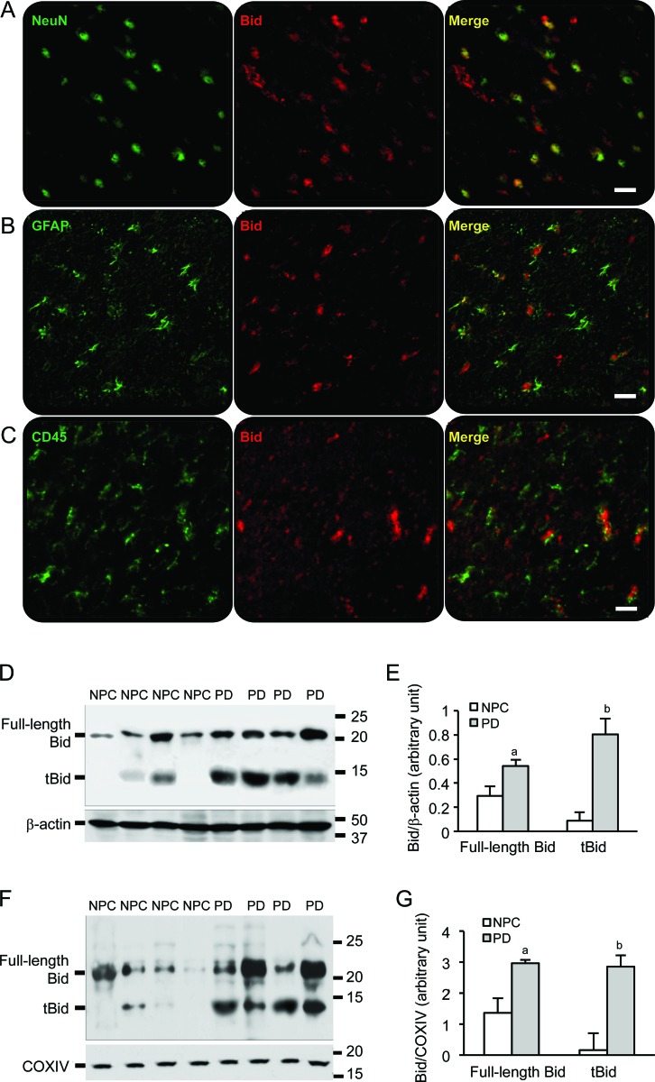
Figure 2. Bid is expressed in neurons and astrocytes, and cleaved Bid is located to the mitochondria in Parkinson disease (PD) brains.
(A) Bid expression was visualized in neurons labeled with antibody against NeuN. (B) Bid expression was found in activated astrocytes identified with antibody against glial fibrillary acid protein. (C) Little Bid expression was found in activated microglia labeled with antibody CD45. Bars: 20 μm. (D) A full-length Bid around 22 kD and the cleaved Bid (tBid) around 14 kD were detected in cytoplasmic fractions of samples from the temporal cortex. (E) Spot density analysis showed increased levels of the full-length Bid and tBid in PD brain samples (ap < 0.05, bp < 0.01). (F) Western blot demonstrated tBid fragments in the mitochondrial fractions from the temporal cortex of PD brains, Cox IV as a mitochondrial loading control. (G) Spot density analysis of increased levels of the full-length Bid and tBid in the mitochondria of PD brain samples (ap < 0.05, bp < 0.01). NPC = nonpathologic control.

