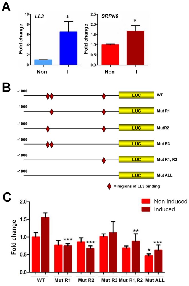
Figure 6. Luciferase expression in a mosquito cell line suggests the involvement of LL3 in the regulation of SRPN6 expression.

Expression of LL3 and SRPN6 mRNAs in response to heat-killed E. cloacae was investigated in hemocyte-like Sua5B cells (A). LL3 and SRPN6 mRNA abundance was determined by qRT-PCR in cells that were non-induced (non) or after exposure to E. cloacae for 6 h (I). Transcript abundance was normalized to that of rpS7 in two independent biological samples and analyzed by the Student's t-test for significance. Asterisks denote significant changes upon bacterial induction (P<0.05). The constructs outlined in (B) were used to assess the ability of LL3 to modulate luciferase expression from a SRPN6 promoter. Red diamonds indicate the locations of LL3-binding sites in the wild type promoter (wt) and their presence/absence in each of the mutated promoter constructs. Individual mutants (R1, R2, or R3) correspond to those sites shown in Figure 5, and were combined to create double (R1,R2) or triple mutants (ALL). (C) Each construct was transfected into Sua5B cells and luciferase expression was measured under basal conditions (non-induced) or upon induction with heat-killed E. cloacae (induced). Expression was normalized to that of the non-induced wild type SRPN6 promoter in triplicate experiments. The values of two biological repeat experiments were pooled. Asterisks denote significant changes when compared to the wild type construct for each treatment (* = P<0.05, ** = P<0.01, or *** P<0.001) as determined by a Two-way ANOVA and Bonferroni post-test.
