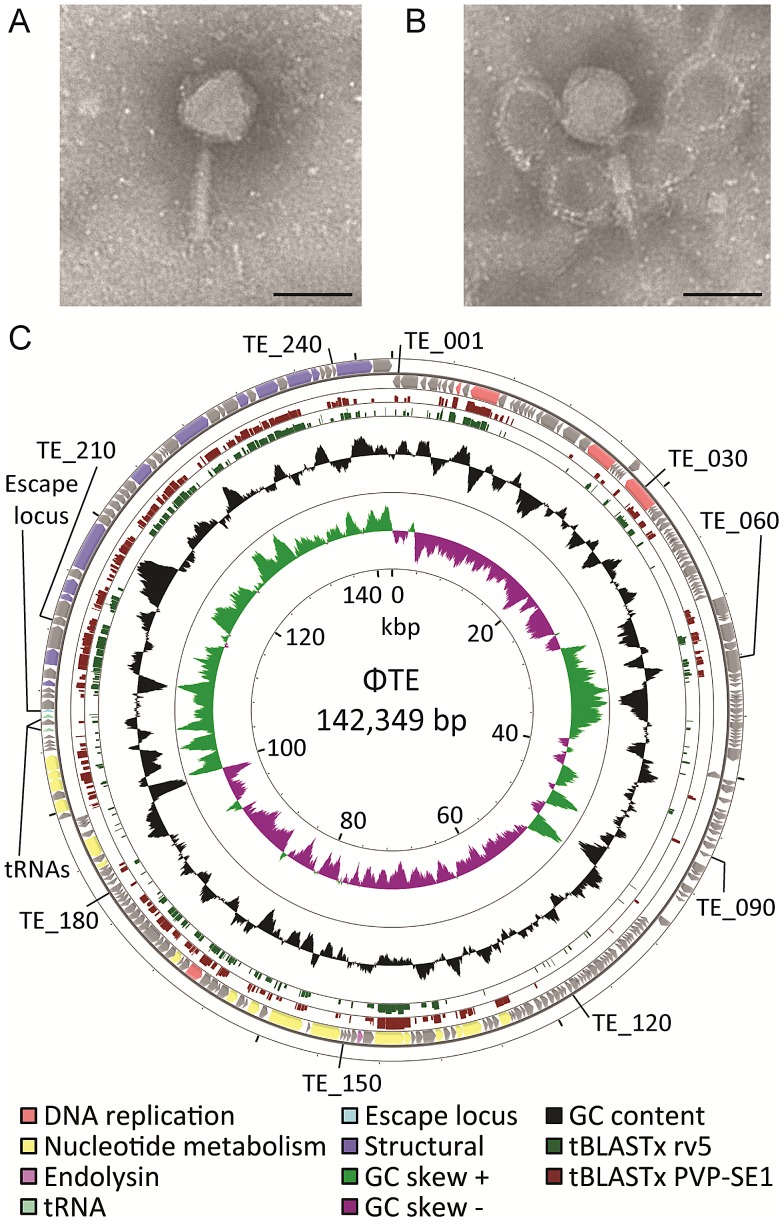
Figure 1. ΦTE morphology and genome overview.
(A–B) Transmission electron micrographs of individual ΦTE virus particles. The tail is fully extended in (A) and contracted in (B). Each scale bar represents 100 nm. (C) Summary of the 142,349 bp circularly-permuted genome of ΦTE, including all ORFs (colour coded to function where possible), two tRNAs and the ncRNA comprising pseudo-ToxI, encoded by the escape locus (Table S2). Selected ΦTE genes are indicated by “TE_x” around the genome, for orientation. GC skew and GC content are shown along with the tBLASTx results against two related phages, coliphage rv5 and Salmonella phage PVP-SE1.

