
Figure 8. Power spectra of membrane potentials for SSVEPs generated with the LCM.

The figure shows (A) the power spectra of membrane potential in layers I, II/III, IV, V, VI and (B) power spectra of the LFP produced by the LCM under intermittent light stimulation. The black lines show power spectra of spontaneous LFPs, and red lines illustrate stimulated LFP power spectra. In (C) an example of LFPs before and after intermittent light stimulation in a single run is also shown. The following parameters were used: 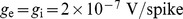 ,
,  for spontaneous activity.