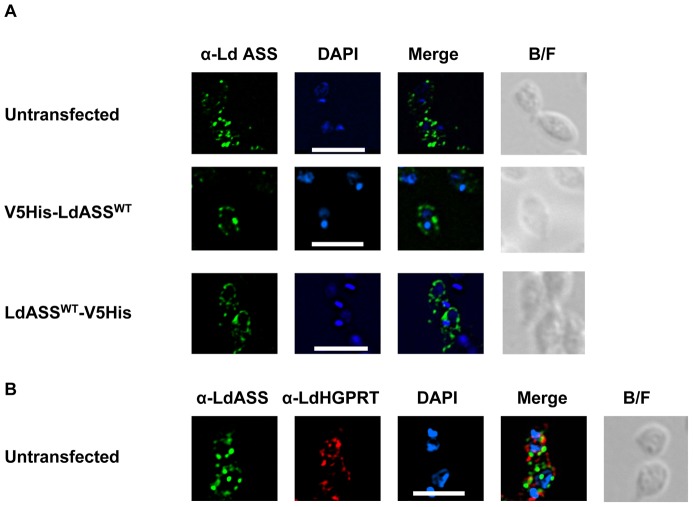
Figure 3. Immunofluorescence analysis of LdASS.
L. donovani amastigote cells were stained with affinity purified antibody against LdASS as primary, and Alexa488-conjugated anti-rabbit IgG (green) as secondary antibodies. Panel A: upper row: untransfected parasites, middle row: V5His-LdASSWT transfected parasites, lower row: LdASSWT-V5His transfected parasites. Panel B: untransfected parasites labeled with biotinylated anti-LdASS (green) and anti-HGPRT antibodies (red). The nuclei and kinetoplast are stained with DAPI (blue). The merge of the images is shown in the column labeled “Merge” in the figure. The right panel shows the bright field images. The white bar represents the scale: 10 µm.

