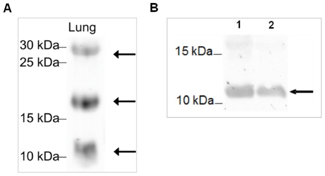
Figure 7. Antibody test by Western blot.

Analyzed was A) lung tissue which is used as positive control for surfactant proteins; B) recombinantly synthesized SP-G protein (not purified) at 28°C (1) and 37°C (2), arrows indicate positive evidence of the surfactant protein G. The proteins extracted from the lung tissue and separated by 15% SDS-PAGE under reducing conditions show distinct bands for SP-G at the theoretically expected molecular weights of 11, 20 and 30 kDA [A]. In case of recombinantly expressed SP-G protein, the antibody detects a distinct band at 11 kDa [B].
