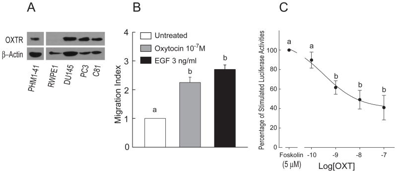
Figure 1.
Oxytocin receptor expression and functions in human prostate cell lines. A, Expression of OXTR was detected with rabbit antibody for OXTR in total cell lysates (50 μg) of RWPE1, DU145, PC3, and C81 cell lines. PHM1-41 expresses endogenous OXTR and was used as a positive control. β-actin was probed on the same blot as sample loading control. B, Treatments with OXT and EGF for 5 h significantly stimulated PC3 cell migration (n=4). C, Twenty-four h after transfection with the cAMP luciferase reporter (pADneo2 C6-BGL), PC3 cells were pretreated with different concentrations of OXT for 10 min before the treatment with forskolin. Luciferase activities were detected 5 h after treatments and expressed as percentage of the forskolin-stimulated luciferase activity (n=3). Data were expressed as Mean ± SEM and analyzed by ANOVA and Duncan’s modified range tests. Significant differences between groups in a given category (P < 0.05) are designated with different lowercase letters.

