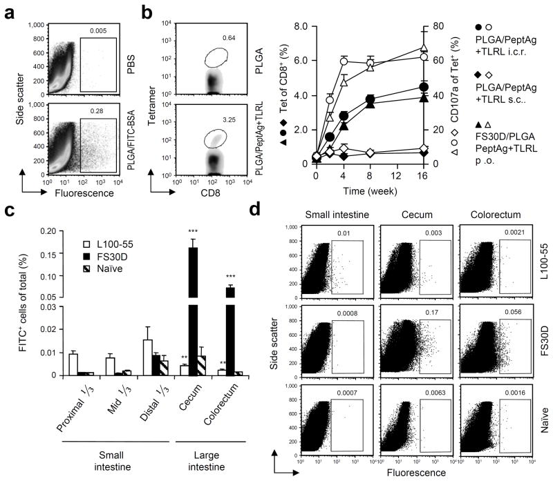
Figure 1.
I.C.R. delivered nanoparticles enter the large intestinal mucosa and orally delivered FS30D/PLGA nanoparticle-releasing microparticles selectively targets the large intestinal mucosa for uptake. (a) Colorectal mucosal uptake of PLGA nanoparticles after i.c.r. delivery of PLGA/FITC-BSA nanoparticles. Cells were isolated from the colorectum 3 days after administration and measured for fluorescence-positive cells. (P < 0.01 between PLGA/FITC-BSA and PBS treated. Representative of three independent experiments, n = 12 15 per group). (b) Induction of antigen-specific colorectal mucosal T cells after i.c.r. delivery of PLGA nanoparticles encapsulating PCLUS3-18IIB and MALP2+poly(I:C)+CpG vaccine (PLGA/PeptAg+TLRL). Three weeks after one immunization, colorectal cells were isolated and measured for P18-I10 specific CD8+ T cells by tetramer staining. P < 0.01 between PLGA/PeptAg+TLRL and PLGA alone. Representative of two independent experiments. n = 10 per group. (c) Gut mucosal uptake of PLGA particles after oral delivery of FS30D/PLGA or L100-55/PLGA. Cells were isolated from the small and large intestine at day 2 for measurement of fluorescence-positive cells. **P < 0.02 on white bar indicates the difference from small intestine. ***P < 0.001 on black bar indicates the difference from the small intestine. n = 9 12 per group. (d) Representative examples of flow cytometry from experiments shown in c.

