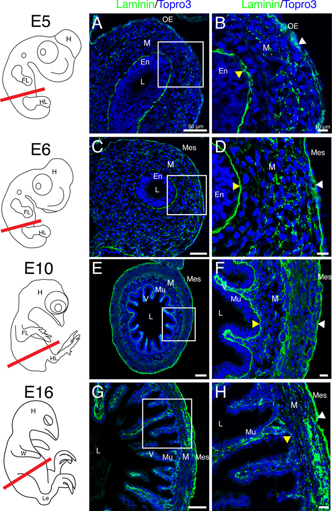
Figure 3. Basement membrane dynamics throughout gut tube closure and mesenchymal differentiation.
Schematics in left column depict quail embryos at the stage isolated and the red line denotes the plane of section. A–B: At E5, the outer epithelial basement membrane appeared dispersed (white arrowhead). Yellow arrowhead denotes the endodermal basement membrane. C–D: At E6, both the outer (white arrowhead) and endodermal (yellow arrowhead) basement membranes were unbroken. E–F: At E10, villi (V) were present and both basement membranes were continuous (arrowheads). G–H: At E16, the mesenchyme was condensed (compare F and H). The outer basement membrane was robust and unbroken (white arrowhead) while the mucosal basement membrane weakly stained with laminin (yellow arrowhead). Scale bars: 50µm (A, C, E, G,) and 10µm (B, D, F, H). En, endoderm; FL, forelimb; H, head; HL, hindlimb; L, lumen; Le, leg; M, mesenchyme; Mes, mesothelium; Mu, mucosa; OE, outer epithelium; V, villi; W, wing.

