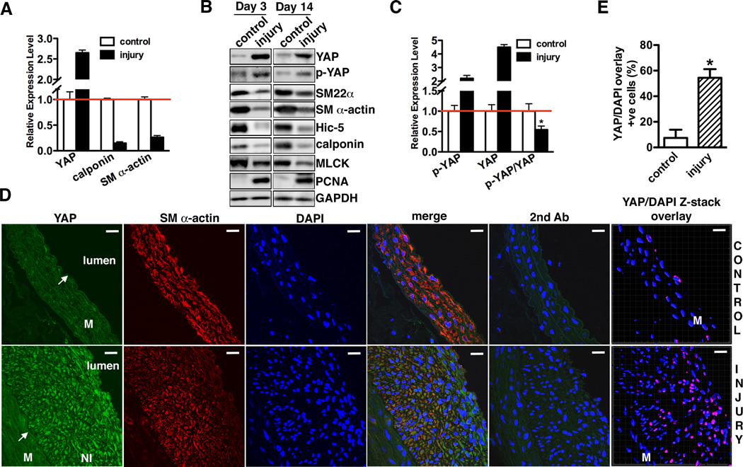
Figure 1. YAP expression is induced in balloon injured rat carotid arteries.
A. qRT-PCR was performed to assess YAP and smooth muscle contractile gene mRNA expression in rat carotid arteries 14 days following balloon injury. Data are expressed relative to control uninjured vessels (set to 1) after normaliztion to RPLP0 as an internal control. N=3. B. 3 or 14 days after rat carotid artery balloon injury control or injured arterial vessels were harvested for Western blotting to assess protein expression as indicated. A representative blot is shown from 4 independent experiments. C. Quantification of immunoblot signals of p-YAP, YAP or p-YAP/YAP shown in “B” of 14 day post injury vessels. *p<0.05. D. 14 days after rat carotid artery balloon injury sections from injured and control arteries were stained for YAP (green) or SM α-actin (red). Samples treated with secondary antibody alone served as negative control. All sections were counter-stained by DAPI to visualize nuclei (blue). Co-localization of YAP and DAPI nuclear staining is shown in purple in control and injured samples by confocal microscope Z-stack scanning and reconstructed by Imaris Bitplane software (far right panel). An arrow points to internal elastic lamina. M: media. NI: neointima. Scale bar, 20um. E. Quantification of the percentage of SMCs that YAP and DAPI staining were co-localized in panel “D” (far right panel). Data were collected from 3 independent experiments. *p<0.05

