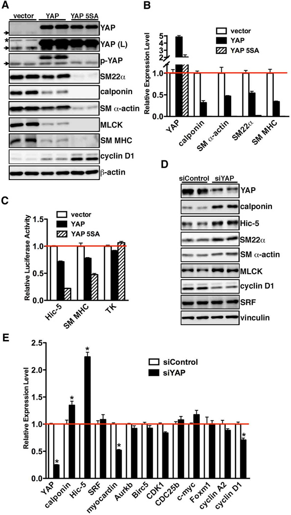
Figure 2. YAP attenuates smooth muscle gene expression.
A. Cultured rat aortic SMCs were transduced with retrovirus encoding wild-type or 5SA mutant YAP, and then harvested for Western blotting or qRT-PCR (B) to evaluate gene expression as indicated. The cells infected with empty retroviral vector served as a control. Arrows point to endogenous YAP or p-YAP signal and an asterisk denotes a non-specific signal after long exposure of the YAP blot (L). As expected, 5SA, a mutant resistant to Hippo kinase, cannot be detected by anti-pYAP (S127) antibody. C. Expression plasmids for WT YAP or YAP 5SA were transfected with either Hic-5, SM MHC or a control minimal thymidine kinase (TK) promoter-luciferase reporter genes into PAC1 SMCs and then promoter activity was measured by dual-luciferase reporter assay. Promoter activity was normalized to a renilla luciferase internal control and expressed relative to vector control transfections (set to 1, red line). Data are presented as mean±SEM of 8 samples. D. Silencing YAP or control RNA duplex was transfected into cultured rat aortic SMCs for 48 hours and then cells were harvested for Western blot or qRT-PCR (E) analysis as indicated. *p<0.05.

