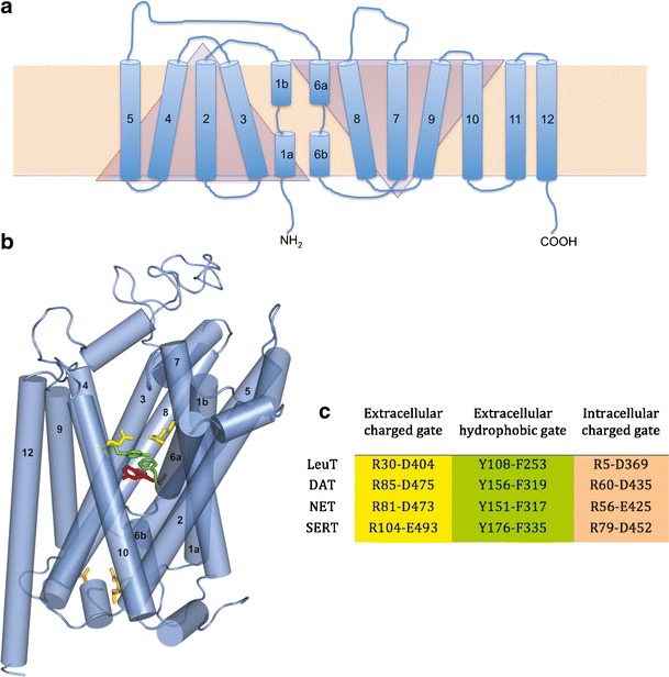
Fig. 1.

LeuT and MAT topology. a 2D representation of LeuT (blue) in the cell membrane bilayer (beige rectangle). TM domains (cylinders) 1–5 and 6–10 are symmetrical but inverted motifs (triangles). b 3D representation of hSERT based on the LeuT (pdb:2A65) crystal structure. The primary substrate binding site (represented by serotonin; magenta) is flanked by gating residues to the extracellular (green, yellow) and intracellular (peach) sides. c LeuT and MAT gating residues, color-coded to match b
