Figure 1.

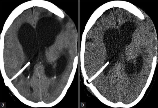
(a) CT head using conventional CT protocol of a patient who presented with a blocked ventriculoperitoneal shunt. (b) CT head using low dose CT protocol of same patient after shunt revision

(a) CT head using conventional CT protocol of a patient who presented with a blocked ventriculoperitoneal shunt. (b) CT head using low dose CT protocol of same patient after shunt revision