Figure 2.

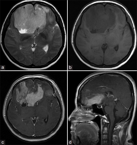
MRI showing bifrontal lesion crossing the midline and displacing the corpus callosum posteriorly (a) axial T2-weighted (b) axial T1-weighted (c) postcontrast axial T1-weighted, and (d) postcontrast sagittal T1-weighted images

MRI showing bifrontal lesion crossing the midline and displacing the corpus callosum posteriorly (a) axial T2-weighted (b) axial T1-weighted (c) postcontrast axial T1-weighted, and (d) postcontrast sagittal T1-weighted images