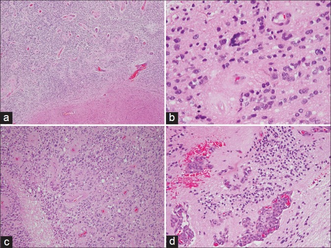
Figure 3.

(a) Low power view showing a cellular tumor with perivascular pseudorosettes and a well demarcated border with the adjacent brain parenchyma (H and E, × 40). (b) An occasional true (ependymal) rosette is identified (H and E, × 400). (c) The tumor is mitotically active and shows areas of necrosis (H and E, × 200). (d) Microvascular proliferation is also present (H and E, × 200)
