Figure 1.

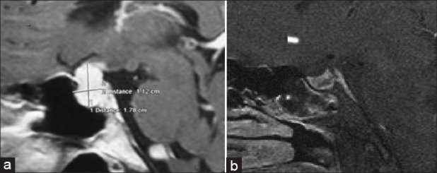
(a) Preoperative image showing 1.78 × 1.12 × 2.2 cm lesion, homogenously hyperintense on T1W postcontrast image, showing thickening of pituitary stalk and dural tail. (b) Postoperatively normal sella with no residual lesion

(a) Preoperative image showing 1.78 × 1.12 × 2.2 cm lesion, homogenously hyperintense on T1W postcontrast image, showing thickening of pituitary stalk and dural tail. (b) Postoperatively normal sella with no residual lesion