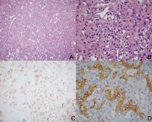
Figure 3.

Histological finding obtained by the biopsy of the lingual lesion. A, B) hematoxylin and eosin appearance of the specimen at different magnification; C, D) same levels of the lesion with a positive focal pattern for CD10 expression (immunohistochemistry).
