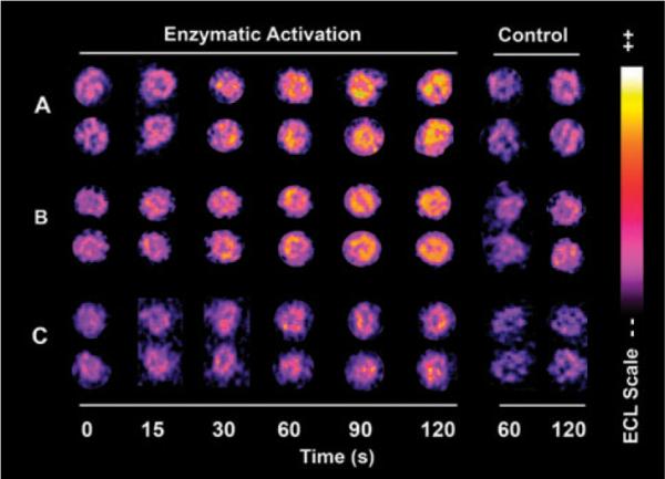
Fig. 1.

Reconstructed ECL array data for (A) spots of RuPVP/DNA/human liver cytosol exposed to 80 μM EDB, 0.5 mM glutathione in 50 mM MES buffer (pH 6.0) for denoted times; (B) spots of RuPVP/DNA/human liver cytosol/human liver microsomes exposed to 100 μM 2-AF, 0.5 mM acetyl coenzyme A, 1.6 mM DTT, 0.5 mM EDTA and NADPH regenerating system in 50 mM MES buffer (pH 6.0); (C) spots of RuPVP/DNA/human liver cytosol exposed to the same enzymatic reaction solution as in (B) without the NADPH regenerating system. Control spots in each panel are exposed to the same concentration of substrates alone.
