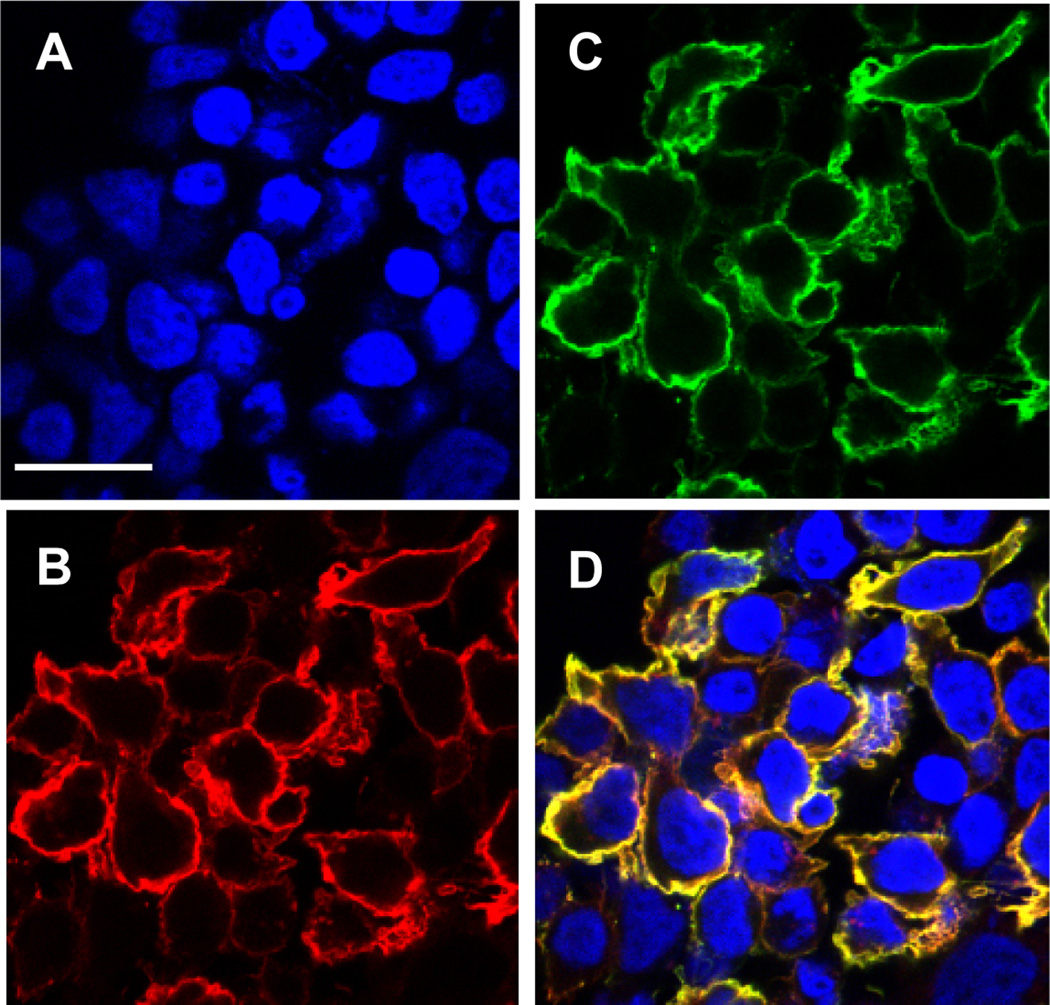
Fig. 3.
Confocal microscopic images of HEK-293T cells displaying scFv. HEK-293T cells transfected with a plasmid directing surface expression of anti-CD22 scFv were grown on cover slips. Transfected cells were fixed with 4,6-diamidino-2-phenylindole nuclear staining (A) followed by detection with biotinylated CD22-Fc (B), and anti-c-myc antibody (C) followed by Steptavidin-Alexa Fluor 594 and anti-mouse IgG-Alexa Fluor 488. (D) shows merged staining patterns. Scale bar = 25 µm. (Originally published in Proceedings of the National Academy of Sciences 103(25):9637–9642, June 20, 2006); copyright 2006 National Academy of Sciences, U.S.A.

