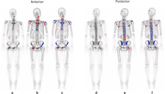
Fig. 1.

A 55-year-old man with lung cancer. Increased radiotracer uptake can mainly be seen in cranial bone, mandible, cervical spine, sternum, and right femur on whole-body scan (a, d). CAD software with European database (b, e) classifies as this patient as having no metastases (ANN 0.29, grade 2). However, CAD software with Japanese database (c, f) correctly classifies as having metastases (ANN 0.87, grade 4). CT imaging shows osteolytic bone metastasis corresponding to increased radiotracer uptake of cranial bone and right femur (not shown)
