
Figure 2.
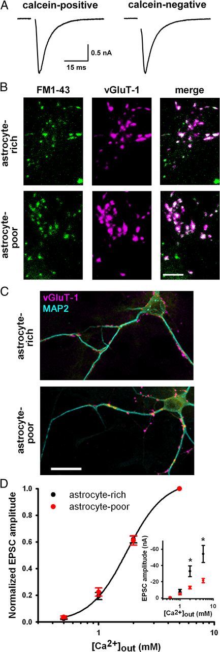
Astrocyte deprivation does not impair basal presynaptic function. A, EPSCs from autaptic neurons in astrocyte-poor cultures from islands with residual live astrocytes (calcein-positive) or no live astrocytes (calcein-negative). B, Images of 10 μm FM1-43FX labeling of presynaptic terminals (green) after 2 min 45 mm KCl and subsequent vGluT-1 immunostaining (magenta) in autaptic neurons from astrocyte-rich and astrocyte-poor cultures (30 min, −20°C, 70% EtOH-fixed astrocytes). Scale bar, 5 μm. C, vGluT-1 (magenta) and MAP2 (cyan) immunostaining in autaptic neurons showing similar density and intensity of presynaptic terminals. Scale bar, 20 μm. Summary results for A–C are given in the Results. D, Calcium concentration–response curves of autaptic EPSC amplitudes from astrocyte-rich or astrocyte-poor cultures normalized for each neuron to values obtained in 5 mm external calcium (n = 21–22). Data from astrocyte-rich cultures were fitted to the Hill equation (EC50 value: 1.8 mm; Hill coefficient: 2.7). Inset, Non-normalized values from the same dataset demonstrating basal depression of EPSCs in astrocyte-poor cultures at higher calcium concentrations. *p < 0.05, Student's unpaired t test, Bonferroni corrected.