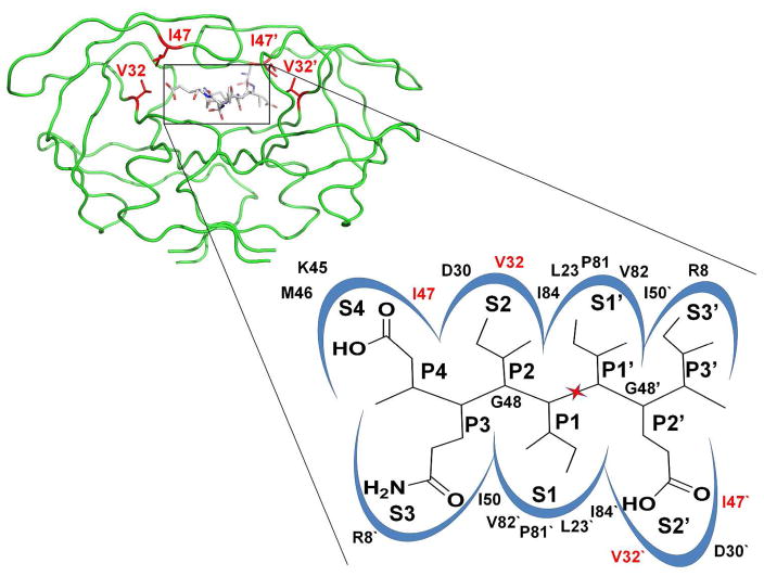
Figure 1.
A) Structure of HIV-1 PR dimer in a green backbone representation. The sites of mutations Val32 and Ile47 are shown as red stick for the side chain atoms in both subunits, with the prime indicating the “second” subunit. The TI peptide is shown in sticks colored by atom type. B) Schematic illustration of the substrate binding site of HIV-1 PR. The peptide DQIIxIEI (P4-P3′) is shown in the S4-S3′ subsites of the PR dimer. The scissile peptide bond is indicated by the red star. PR residues contributing to the binding site are indicated.

