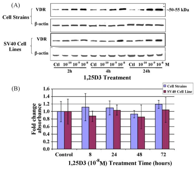
Fig. 1.
The concentration-dependent effect of 1,25D3 on VDR protein levels within dolphin skin cells. (A) VDR protein levels were assessed by Western blots after solvent (ctl), 10−10, 10−9, or 10−8 M concentrations of a 1,25D3 compound were administered to dolphin cell strains and SV40 cell lines for 2, 4, or 24 h. The same blot was stripped and reprobed with β-actin. (B) MTT assays measured cell viability as influenced by 10−8 M 1,25D3 treatment for the indicated times. Columns represent the average fold change in absorbance and bars the standard deviation among eight replicates. No significant differences in cell viability were detected within either the cell strains or the SV40 cell lines (one-way ANOVA, p < 0.05).

