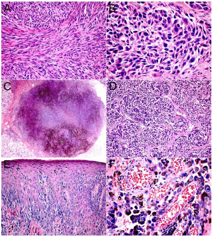
Figure 2.
Histological aspects of primary oral melanoma (H&E). A. Oral melanoma of the hard palate composed mainly by amelanotic spindle cells (x200). B. Oral melanoma of the upper gingiva formed by epithelioid and plasmacytoid neoplastic melanocytes (x400). C. Solid OM showing nodular pattern of neoplastic melanocytic cells (x25). D. Epithelioid and plasmacytoid neoplastic melanocytes arranged in organoid pattern (x100). E. In situ lesion with neoplastic melanocytes arranged in pagetoid pattern (x100). F. Oral melanoma of the hard palate showing perivascular invasion (x400).

