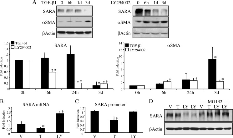
FIGURE 3.
The mechanism of down-regulation of SARA expression differs between PI3K inhibition and TGF-β1. A, HKC were treated with either 2 ng/ml TGF-β1 (left panels) or 20 μm LY294002 (right panels) for the indicated time periods prior to lysis for Western blot. β-Actin is included as a control for loading. As demonstrated in graphs of triplicate experiments, LY294002 reduces SARA (left graph) and increases αSMA (right graph) at all treatment times compared with control (*, for SARA: 6 h, p = 0.0001; 24 h, p = 0.0001; 3 days, p = 0.0001. For αSMA, 6 h, p = 0.002; 24 h, p = 0.0005; 3 days, p = 0.0012). However, TGF-β1 only causes significant changes in SARA and αSMA at 3 days (3d). (*, for SARA, 3 days, p < 0.0001; and for αSMA, p = <0.0001). B, HKC treated for 48 h with either DMSO vehicle (V), 2 ng/ml TGF-β1 (T), or 20 μm LY294002 (LY) were assayed for mRNA expression of SARA via quantitative RT-PCR. TGF-β1 reduced SARA mRNA expression (*, p = 0.0438), but LY294002 enhanced it (*, p = 0.0001). C, HKC were transfected with the putative SARA promoter/luciferase reporter construct for 3 h prior to treatment as in B with TGF-β1 or LY294002 for an additional 48 h. TGF-β1 reduces SARA promoter activity (*, p = 0.002), LY294002 does not (p = 0.8164). D, HKC were pretreated with 10 μm MG-132 for 1 h prior to 24 h treatment with either vehicle (V), TGF-β1 (T), or LY294002 (LY). β-Actin is included as a control for loading.

