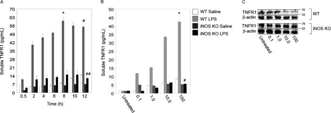
FIGURE 2.
iNOS is essential for TNFR1 shedding into cell culture medium. A, LPS induces TNFR1 shedding into the extracellular medium. ELISA was used to measure the amount of TNFR1 in medium, and raw optical density values were converted to soluble TNFR1 concentrations via a standard curve followed by normalization to PBS-treated hepatocytes. At 8 h, an 18-fold increase in optical density was measured for WT 100 ng/ml LPS-treated cells (*, p < 0.01; 0.5 versus 8 h by paired t test; n = 4–6 per time point; * represents mean ± S.E.). At 12 h, a 21-fold increase in optical density was measured for both WT and iNOS knock-out (KO) hepatocytes (*, p < 0.01; WT versus iNOS KO at 12 h by paired t test; n = 4–6 per time point; * represents mean ± S.E.). B, TNFR1 released into the medium from cells subjected to LPS at different concentrations (0.1–100 ng/ml) and saline (PBS) as a control. Culture medium was sampled, and raw optical density was normalized to the signal from untreated (medium only) cells. A 20-fold increase in soluble TNFR1 was noted after 12 h of 100 ng/ml LPS-treated cells compared with untreated cells (*, p < 0.01; n = 4–6 per condition; * and # represent mean ± S.E.), and a 13-fold increase was noted in soluble TNFR1 in WT hepatocytes versus iNOS KO hepatocytes (#, p < 0.01; n = 4–6 per condition). C, WT and iNOS KO hepatocytes were treated with different concentrations of LPS and analyzed by Western blot at 12 h after treatment. Error bars indicate mean ± S.E.

