FIGURE 2.
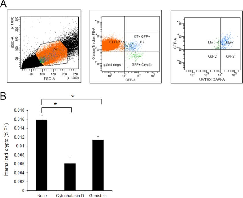
C. neoformans internalization into HBMEC involves host actin cytoskeleton rearrangement. Cryptococcal internalization was determined using flow cytometry with GFP-expressing Cryptococcus cells. HBMEC monolayers were labeled with CellTracker CMTMR Orange (red) and incubated with GFP-expressing cryptococci (crypto, green) for 3 h. The HBMEC were washed with PBS to remove unbound fungi followed by incubation with trypsin/EDTA to dissociate bound fungi from HBMEC as well as to collect HBMEC. The cell mixture was subsequently stained with Uvitex-2B (blue). A, the mixed population of HBMEC and fungal cells was analyzed by flow cytometry using triple filter sets. P1 region (left) of total population was analyzed with red (Orange Tracker) and green (GFP) filters, and then the population showing double-positive (P2 region, middle) was further analyzed with blue (DAPI) filter. The population exhibiting Green+/Red+/Blue− was determined as the HBMEC with internalized fungal cells (right). B, a cryptococcal internalization assay was repeated in the presence of inhibitors. The percentages of HBMEC with internalized fungal cells among the P1 region were plotted. These experiments were repeated twice independently. *, p < 0.001.

