FIGURE 1.
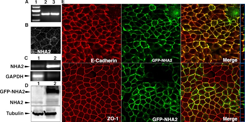
NHA2 expression and localization in MDCK cells. A, reverse transcription-PCR of NHA2 in MDCK cells (lane 1), control plasmid (lane 2). B, immunostaining of endogenous NHA2 using anti-NHA2 antibody. C, reverse transcription-PCR comparing NHA2 expression from control cells (top panel, lane 1) with stably transfected cells overexpressing NHA2-GFP (top panel, lane 2); bottom panel: GAPDH controls. D, Western blot of total protein from control cells (top panel, lane 1) and stably transfected clone overexpressing NHA2-GFP (top panel, lane 2) using anti-NHA2 and anti-GFP antibodies; bottom panel: tubulin controls. E, confocal microscopic images; top panel: NHA2-GFP (green) colocalizes with basolateral marker E-Cadherin (red); bottom panel: NHA2-GFP (green) is found basolateral to the tight junction marker ZO-1 (red). Blue boxes on the right show an orthogonal view of a slice from the confocal image.

