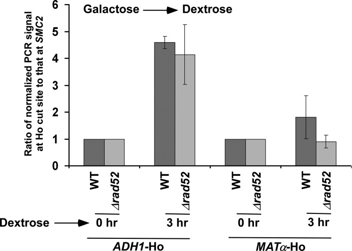
FIGURE 4.
Analysis of DSB repair at the ADH1 and MATα loci in the presence and absence of Rad52p. Both the wild-type (PCY23) and Δrad52 (PCY36) strains were grown as in Fig. 3B. Genomic DNAs from these yeast cultures were prepared and analyzed by PCR for DSBs at the ADH1 and MATα loci. A specific region within SMC2 was amplified as control, using the same genomic DNAs. The PCR signals at 0 h time point for the ADH1, MATα and SMC2 loci were set to 100, and the PCR signals at 3 h were normalized with respect to 100. The ratios of normalized PCR signals at 3 h at the ADH1 and MATα Ho cut sites to that at SMC2 are plotted in the form of a histogram. A ratio that is greater than 1 would indicate the DSB repair.

