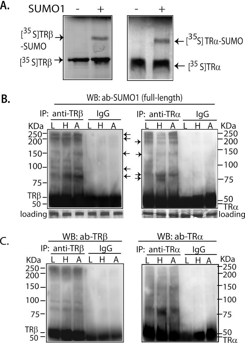
FIGURE 1.
In vitro and in vivo sumoylation of TR. A, [35S]TRα and -TRβ were incubated with E1, UBC9, and SUMO1 in sumoylation buffer at 37 °C for 1 h and then separated by 10% SDS-PAGE. B, mouse liver (L), heart (H), and white adipose (A) tissues were utilized for IP with anti-TRα (rabbit IgG) and anti-TRβ (mouse IgG) antibodies. In WB, anti-full-length SUMO1 was used to detect sumoylation. The membranes shown in B were then stripped, checked by chemiluminescent exposure to confirm the absence of residual activity, and then incubated with anti-TRβ (rabbit IgG) and anti-TRα (rabbit IgG) antibodies (ab; C) to confirm that the identified TR-SUMO bands contained TR. IgG was used as nonspecific control. Arrows indicate the location of TR-SUMO complexes.

