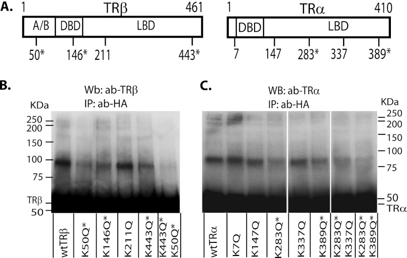
FIGURE 3.
Identification of the sumoylation sites in TRα and TRβ. A, predicted sumoylation sites based on amino acid sequence in TRα and TRβ, with an asterisk showing confirmed sites based on mutation analysis. HepG2 cells were transfected with vectors expressing SUMO1-HA (B; for TRβ) or SUMO3-HA (C; for TRα) and wild type or mutant TRs as shown. Cells were treated with T3 (50 nm) for 4 h for TRβ sumoylation, but without T3 for TRα sumoylation. The cell lysate was immunoprecipitated with an anti-HA antibody (ab) to detect SUMO1- or SUMO3-HA, and Western blot was performed with TRβ (B) or TRα (C) antibody. *, confirmed sumoylation sites in TRs. A/B, activation domain; DBD, DNA binding domain; LDB, ligand binding domain.

