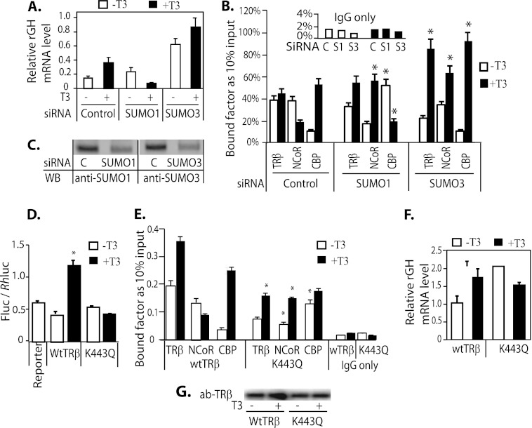
FIGURE 6.
The influence of SUMO1 and SUMO3 expression on endogenous rGH mRNA expression and recruitment of TRβ and cofactors to the TRE. A, GH3 cells were transfected with siRNA SUMO1 or SUMO3. Three days after transfection, the medium was changed to serum-replaced medium, and cells were allowed to grow for 16 h. Cells were treated with or without T3 (50 nm) for 4 h prior to isolating RNA. The endogenous rGH mRNA expression in GH3 cells was detected by q-PCR. C, control. B, ChIP assays were performed using GH3 cells transfected with TRβ and siRNA SUMO1 or siRNA SUMO3 and antibodies (ab) anti-TRβ, anti-NCoR, and anti-CBP. C, WB analysis of SUMO1 and -3 knockdown. D, the rGH TRE-luc reporter activity was analyzed in TRβ- and TRβ K443Q-transfected GH3 cells with or without addition of 50 nm T3. E, ChIP assay of TR binding to rGH TRE and interaction with NCoR and CBP. GH3 cells were transfected with TRβ and TRβ K443Q and treated with or without T3 as described in A. Antibodies used in ChIP assay were anti-TRβ, anti-NCoR, and anti-CBP. The specific region of the rGH TRE was q-PCR quantified (see Table 1 for primers). F, endogenous rGH mRNA expression in the presence of transfected TRβ or TRβ K443Q. T3 treatment is the same as described in A. G, WB shows the protein level of TRβ and TRβ K443Q in transfected cells from the experiment shown in E. *, indicate p < 0.05 compared with controls.

