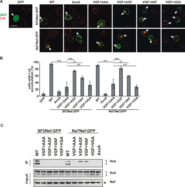
Figure 6.
The VGF region is important for the interaction of Nef with Lck and Hck. (A) Jurkat TAg cells were transfected arrows with WT, AxxA, VGF→AAA, VGF→AGF, VGF→VAF or VGF→VGA mutant Nef.GFP fusion proteins, plated onto poly-L-lysine (PLL)–coated cover glasses, fixed and stained for Lck (red): wild-type but not AxxA Nef induces Lck accumulation (arrow heads), Images shown are representative for all cells analyzed. Scale bar = 10 μm (B) Frequency of the cells from cultures as shown in panel A that show Lck accumulation. Values are the means of 3 independent experiments, and error bars represent SD from the mean; ≥ 100 cells were analyzed per transfection, ** indicates P < 0.001 and *** indicates P < 0.0001. (C) Western blot of lysates of 293 T cells co-expressing WT, VGF→AAA, VGF→AGF, VGF→VAF, VGF→VGA or AxxA SF2 or NA-7 Nef.GFP fusion proteins (all similar fraction of cells positive for GFP) or GFP only together with Hck. Lower panels (input) show staining for Nef (arrow) and Hck; upper panel shows anti-GFP immunoprecipitation (IP) stained for Hck. Interaction of Nef with Hck was detected as the presence of Hck isoforms in the immunoprecipitate.

