
Figure 4. Acquired epilepsy is associated with alterations in [Ca2+]i and Ca2+ homeostatic mechanisms 24 h post-in vitro SE.
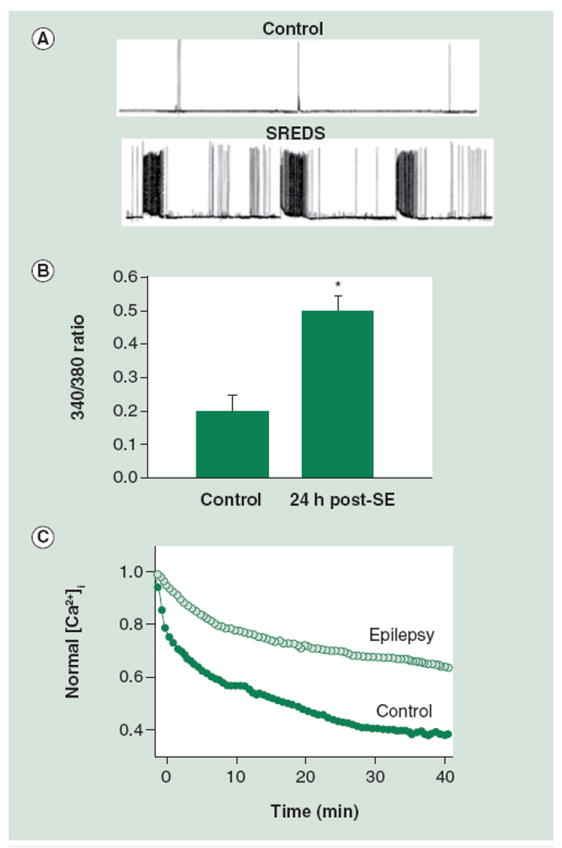
(A) Electrophysiological traces using current-clamp technique demonstrate baseline activity in control neurons and SREDs in neurons that had been treated in low Mg2+ for 3 h and then returned to normal maintenance media. Resting membrane potential = -61.5 mV; duration of shown trace = 30 min. (B) Bar graph comparing 340/380 ratio values in control versus SE groups 24 h after treatment with Mg2+-containing buffer solution or low Mg2+, respectively. Neurons in the SE group exhibit a 340/380 ratio of 0.5 ± 0.08 (mean ratio ± standard error of the mean). The ratio from control group neurons is significantly lower at 0.20 ± 0.05. (C) Glutamate stimulation causes increased [Ca2+]i. Peak ratios following glutamate are normalized to 1.0. Recovery from this challenge is hindered in epileptic neurons when compared with controls.
[Ca2+]i: Intracellular Ca2+ concentration; SE: Status epilepticus; SRED: Spontaneous, recurrent epileptiform discharge.
(A) reproduced with permission from [83] and (B) reproduced with permission from [37].