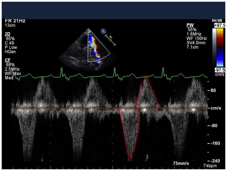
Figure 1.

Pulsed wave Doppler in the main pulmonary artery in a patient with repaired tetralogy of Fallot and residual pulmonary regurgitation. Diastolic (above the baseline) and systolic (below the baseline) flows were traced (shown in red) to obtain the DSTVI. In this example, DSTVI= 0.612, corresponding to moderate-severe pulmonary regurgitation by CMR (RF = 47%).
