
Figure 4. Nogo-A-specific antibody enhanced corticospinal axon sprouting.
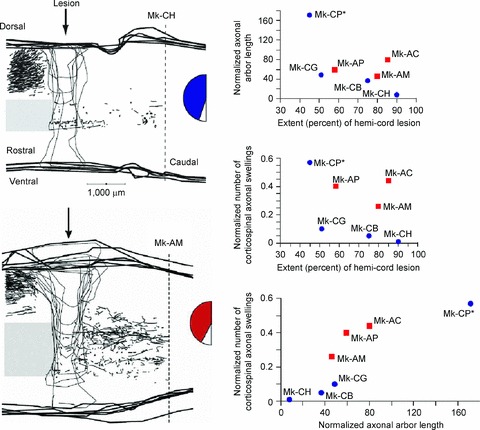
Corticospinal axons were labelled using biotinylated dextran amine (BDA) injections in the contralateral motor cortex. BDA-labelled axonal arbors caudal to the lesion were more numerous in monkeys treated with Nogo-A-specific antibody (Mk-AM; left, bottom) than with control antibody (Mk-CH; left, top). Seven monkeys were used to determine the normalized cumulative corticospinal axonal arbor length (in mm; right, top) and the normalized number of axonal swellings by corticospinal axons (i.e. putative re-established contacts with interneurons or motoneurons; right, middle), which were plotted as a function of lesion extent. Monkey Mk-CP had profuse sprouting but was incompletely lesioned. The number of corticospinal axonal swellings was also plotted as a function of the cumulative corticospinal axonal arbor length (in mm) (right, bottom). In all graphs, blue circles represent control antibody-treated monkeys and red squares represent Nogo-A-specific antibody-treated monkeys. The extent of the blue and red zones in the semicircular figures represents the extent of the hemicord lesion. Figure modified from Fig. 2 in Freund et al. 2006.