
Figure 5. Cocaine treatment increases putative apposition of excitatory VGLUT2 puncta.
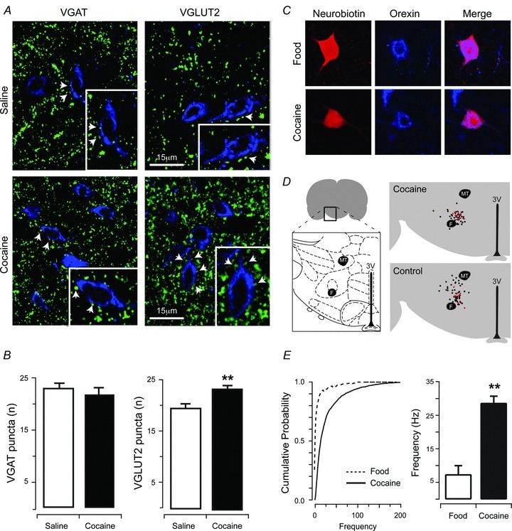
A, dual-labelling for VGAT and orexin (left), or VGLUT2 and orexin (right). Upper panels show saline-treated animals and lower panels show cocaine-treated animals. B, cocaine exposure did not increase putative apposition of inhibitory VGAT puncta onto orexin neurones. In contrast, cocaine exposure significantly increased putative apposition of excitatory VGLUT2 puncta onto orexin neurones (**P < 0.01). C, image panels show immunolabelling for neurobiotin (left), or orexin (middle), and merged pictures (right) from a recorded neurone confirming that population of recordings were from orexin positive neurones. Upper panels are from food-trained controls and lower panels are from cocaine-trained rats. D, schematic diagram showing the collated locations of all recordings in cocaine-exposed and control animals, with red dots indicating confirmed orexin positive recordings (Bregma 2.76–Bregma 3.24). Note that recovery of neurobiotin and orexin labelling was only performed for cocaine self-administration experiments. E, cumulative probability of orexin neurones (from representative recordings, left) and bar plots (group data, right) of mEPSC frequency highlight a significant increase in events recorded from cocaine- compared to food-trained rats (**P < 0.01).