
Figure 5. CRF effects are mediated by CRF1.
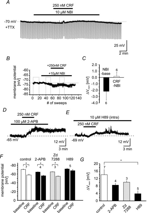
A, example recording of an individual NPS-EGFP neuron in current-clamp mode at a membrane potential of −70 mV. The recording was done in the presence of TTX. Hyperpolarizing currents (−60 pA; 500 ms duration) were injected to monitor the input resistance. Bars indicate the duration of NBI27914 (10 μm) and CRF (250 nm) application. B, scatter plot of the mean membrane potential vs. number of recorded sweeps (sweep duration: 10 s). Dashed boxes indicate the time intervals taken for quantification (baseline; NBI-application; NBI + CRF application). C, quantification of the change in membrane potential induced by NBI (NBI – baseline) or by CRF (CRF – NBI). D, representative current-clamp recording of a single NPS-EGFP neuron. CRF was applied in the presence of 100 μm 2-APB. E, example of a current-clamp recording at −69 mV membrane potential. The intracellular recording solution contained the PKA antagonist H89 (10 μm). Note the reduced depolarization induced by CRF in presence of H89. F, quantification of the membrane potential of NPS-EGFP neurons during baseline conditions and in the presence of CRF during control or in combination with 2-APB, ZD 7288 and H89. G, quantification of the CRF-induced depolarization during control or in the presence of 2-APB, ZD 7288 and H89.