
Figure 6. D1 and A1 adenosine receptors modulate presynaptic activity in fluorescent cells from Drd1-EGFP mice.
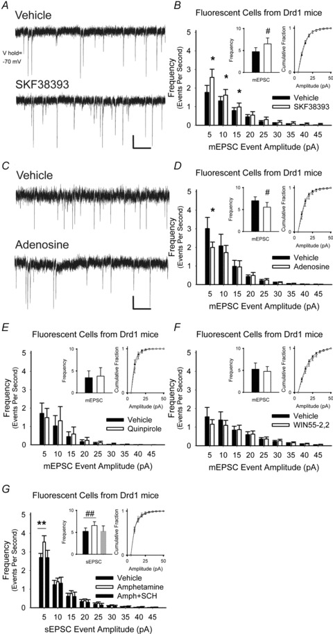
A, representative mEPSCs in D1R+ cells (upper trace) increased 5 min after exposure to the D1R agonist SKF38393 (lower trace). B, SKF38393 increased the frequency of mEPSCs (inset, left) by boosting the number of high-frequency, low-amplitude spontaneous inward currents while having no effect on the cumulative distributions of mEPSC amplitudes (inset, right). For panels B and D–G, *P < 0.05, **P < 0.01, Student's t test with Bonferroni adjustment and #P < 0.05, ##P < 0.01, Student's paired t test. C, representative mEPSCs in D1R+ cells (upper trace), decreased after exposure to adenosine (lower trace). D, adenosine reduced the frequency of low-amplitude mEPSCs but had no effect on the distribution of mEPSC amplitudes. E, the D2R agonist quinpirole did not alter the frequency, the frequency distribution or the amplitude distribution of mEPSCs in D1R+ cells. F, the CB1R agonist WIN55-2,2 also had no effect on the mEPSC frequency, the frequency distribution or the amplitude distribution in D1R-expressing cells. G, amphetamine increased in frequency, but not amplitude distribution, of sEPSCs D1R+ cells. Excitation by amphetamine was blocked by the D1R antagonist SCH23390 (SCH). Bar in panels A and C, 5 pA, 0.5 s.