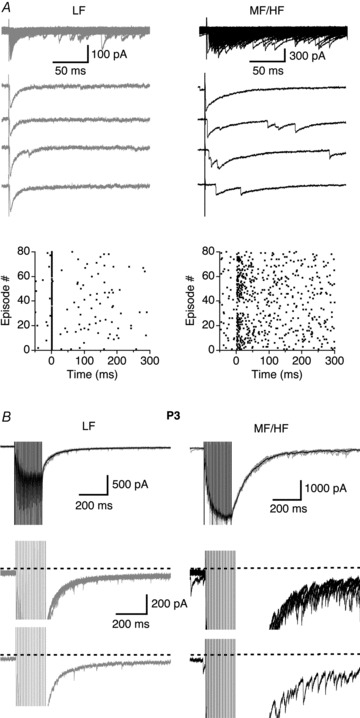
Figure 5. Prominent asynchronous GABA release was present in MF/HF neurons under minimal stimulation conditions and in more mature neurons.

A, superimposed IPSCs (top) and four individual traces for each neuron elicited by single-pulse stimuli under minimal stimulation conditions. The LF neuron exhibited a stimulus time-locked response representing strong synchronous release, whereas in the MF/HF neuron IPSCs were more scattered in their timing of occurrence. The zero time point in the raster plots indicates the onset of the electrical stimulation. B, in P3 chicks, more prominent asynchronous release events seemed to be present in MF/HF neurons. Upper row, superimposed IPSCs (grey) elicited by train stimulations (100 Hz, 20 pulses) with the average trace shown in black. Middle and lower rows, superimposed IPSCs and one IPSC trace respectively from the upper row shown at an enlarged amplitude scale.
